Juan M Pérez1, Sara Robledo2,3, Wilson Cardona1, Fernando Alzate4, Diana Muñoz3, Angie Herrera1
1Quimica de Plantas Colombianas, Instituto de Química, Facultad de Ciencias Exactas y Naturales; 2Programa de Estudio y Control de Enfermedades Tropicales (PECET), Sede de Investigación Universitaria, SIU; 3CIDEPRO-Center for Development of Products against Tropical Diseases; 4Grupo de Estudios Botánicos, Instituto de Biología, Facultad de Ciencias Exactas y Naturales, Universidad de Antioquia UdeA, Calle 70 No. 52–21, A.A 1226, Medellín, Colombia.For correspondence:- Wilson Cardona Email:
Received: 11 October 2015 Accepted: 12 April 2016 Published: 27 May 2016
Citation: Pérez JM, Robledo S, Cardona W, Alzate F, Muñoz D, Herrera A. Leishmanicidal and cytotoxic activity of extracts and saponins from Ilex laurina (Aquifoliaceae). Trop J Pharm Res 2016; 15(5):973-979 doi: 10.4314/tjpr.v15i5.11
© 2016 The authors.
This is an Open Access article that uses a funding model which does not charge readers or their institutions for access and distributed under the terms of the Creative Commons Attribution License (http://creativecommons.org/licenses/by/4.0) and the Budapest Open Access Initiative (http://www.budapestopenaccessinitiative.org/read), which permit unrestricted use, distribution, and reproduction in any medium, provided the original work is properly credited..
Purpose: To evaluate the leishmanicidal and cytotoxic activity of alcohol and non-alcohol extracts and saponins from Ilex laurina.
Methods: Extracts were obtained by percolation with solvents of different polarities: hexane, dichloromethane, ethyl acetate and ethanol. The ethyl acetate extract was subjected to silica gel column chromatography eluting with a step gradient of dichloromethane-methanol. All products were evaluated in vitro for leishmanicidal activity against amastigotes of leishmania panamensis and cytotoxicity on U-937 cells.
Results: Two saponins were isolated from the ethyl acetate extract. The ethyl acetate extract showed high leishmanicidal activity against intracellular amastigotes of L. panamensis (EC50, 7.5 ± 1.5 µg/mL) and low activity against axenic amastigotes (EC50, 52.8 ±1.6 µg/mL); this extract showed also high cytotoxicity (LC50, 57.7 ± 12.1 µg/mL). Saponin 2 exhibited high activity against intracellular amastigotes (EC50, 5.9 ± 0.5 µg/mL) but also showed high cytotoxicity on U-937 cells (EC50, 25.7 ± 6.1 µg/mL). This compound showed similar leishmanicidal activity and cytotoxicity to meglumine antimoniate and amphotericin B, respectively, drugs currently used for the treatment of leishmaniasis.
Conclusions: Based on these results, Ilex laurina is a potential source of compounds that can lead to the development of new therapeutic alternatives against leishmaniasis.
Introduction
Protozoan parasites are responsible for some of the most common and devastating diseases affecting humans and other mammals [1]. Among these diseases is leishmaniasis, which causes the most deaths after malaria [2], the reason why it has become a priority for the World Health Organization (WHO) [2].
Cutaneous leishmaniasis is endemic in the tropics and sub-tropics. It is often referred to as a group of diseases because of the broad spectrum of clinical manifestations, which range from small cutaneous nodules to gross mucosal tissue destruction. In America, cutaneous leishmaniasis can be caused by a multitude of Leishmania species, members of the Vianna subgenus, such as L. (V.) panamensis, L. (V.). braziliensis, and L. (V.). guayanensis, and members of the Leishmania subgenus, such as L. (L.) amazonensis and L. (L.) mexicana [3]. Leishmaniasis caused by these Leishmania species are associated with recurrences or relapses and lesions are hard to heal in absence of treatment with specific drugs.
Classical antileishmanial drugs, pentavalent antimonials (meglumine antimoniate and sodium stibogluconate), pentamidine isothianate and miltefosine, show high toxicity, important side effects and are largely no longer effective because of the emergence of resistance in the parasites, thus becoming a major public health problem [4,5]. The lack of an effective leishmanicidal drug has evoked a renewed interest in medicinal plants as sources of new chemotherapeutic compounds that are more effective and have fewer side effects [6-9].
In order to find new drugs to combat this disease, we have studied extracts of Ilex laurina, a native Colombian plant that is distributed in the country in the north of Central and Western mountain ranges, from 1600 to 2900 m [10]. The Ilex genus belongs to the Aquifoliaceae family, comprising nearly 400 species of global distribution [11]. In Colombia, Ilex genus involves 35 species. I. paraguariensis or “yerba mate” is the most commercialized plant of South America because infusions of aerial parts show a variety of biological properties including antioxidant activity [12,13], cardiovascular effects [14,15], anti-parkinsonian effect [16], hypocholeste-rolemic, hepatoprotective [12], stimulant of the central nervous system and diuretic [17]. Different Ilex species are used as tea for protection against heart and liver diseases, brain dysfunction, and maintenance of proper body weight [18]. Tea of Ilex guayusa (named “guyasa”) is used in southern Colombia (Amazon) and northern Ecuador as mild stimulant [19]. Antioxidant, antiproliferative and apoptotic activities of Ilex laurina infusion have also been reported [20].
Based on the broad spectrum of biological activities that have been reported for Ilex species and the need for new therapies against leishmaniasis, this study aimed at evaluating the leishmanicidal and cytotoxic activity of extracts and secondary metabolites from I. laurina.
Methods
Plant materials
Leaves of I. laurina were collected during June 2013 in the village of Santa Elena, municipality of Medellín (Antioquia, Colombia) and identified by Dr. Fernando Alzate (Biology Institute, University of Antioquia, Medellin, Colombia). A voucher specimen (no. Alzate-50622) was kept at University of Antioquia Herbarium.
Extraction and isolation
Powdered leaves (0.7 kg) of I. laurina were extracted successively with hexane, dichloromethane, ethyl acetate, and ethanol (10L each) in a percolator at room temperature and concentrated in vacuum to give the corresponding yields (66.6g [8.9 %], 124.3g [16.6 %], 107.3g [14.3 %] and 128.4g [17.1 %], respectively).
The ethyl acetate extract was subjected to silica gel column chromatography (5 × 80 cm) eluting with a step gradient of dichloromethane-methanol (100:0, 90:10, 80:20, 70:30, 60:40, 50:50, 40:60, 30:70, 20:80,10:90, 0:100, each 500 mL), to obtain 10 fractions (F1 - F10) collected on the basis of their TLC profiles (dichloromethane-methanol-acetic acid 80:15:5). Fractions F4 and F8 were recognized as the most interesting ones, due to the appearance of black spots after spraying with universal reagent. Compounds 1 (30 mg, 5.8 %) was isolated from F8 and compound 2 (35 mg, 3.9 %) from F4, by preparative TLC using dichloromethane-methanol-acetic acid (80:15:5) mixture.
Biological assays
The extracts and compounds were subjected to in vitro cytotoxicity on mammalian cells and leishmanicidal activity on axenic and intracellular amastigotes of L. (V.) panamensis.
In vitro cytotoxic activity in mammalian cells
The cytotoxicity of the products was assessed based on the viability of the human promonocytic cell line U937 (ATCC CRL-1593.2TM) evaluated by the MTT (3 - (4, 5-dimethylthiazol-2-yl) - 2, 5-diphenyltetrazolium bromide) method as described previously [21, 22]. Briefly, into each well of a 96-well cell-culture dishes were dispensed 100,000 cells/100 μL in RPMI-1640 supplemented with 10 % FBS and 100 uL of the corresponding concentrations of the extracts and saponins. Six double serial diluted concentrations were evaluated starting at 200 µg/mL. The cells were incubated at 37 ºC with 5 % CO2 for 72 h in the presence of the extracts and saponins, and then the effect was determined by measuring the activity of the mitochondrial dehydrogenase by adding 10 µL/well of MTT solution (0.5 mg/mL) and incubating at 37 ºC for 3 h. The reaction was stopped by adding a 50 % isopropanol solution with 10 % sodium dodecyl sulfate for 30 min. Cell growth was determined based on the quantity of formazan produced, which was measured at 570 nm in a benchmark reader plate spectrophotometer (Bio-Rad Hercules, CA, USA). Cells cultured in the absence of products were used as cell growth controls, while cells cultured in presence of meglumine antimoniate and amphotericin B were used as cytotoxicity controls. Each concentration was tested in triplicate in two independent experiments.
Cytotoxicity was determined in terms of cell viability and cell growth inhibition obtained for each compound, amphotericin B or medium alone. Percentages of viability were calculated using Eq 1.
Viability (%) = (At/Ac)100 …………………. (1)
where At is the absorbance of treated cells and Ac (i.e., absorbance of control cells) corresponds to 100 % viability.
Cell growth inhibition was calculated as in Eq 2.
Cell growth inhibition (%) = 100 – viability (%) …… (2)
The results are expressed as 50 % lethal concentration (LC50), i.e., the concentration of drug that gives half-maximal inhibition of cell growth. LC50 was calculated by Probit method [23]. The cytotoxicity of each product was graded according to the LC50 value, based on this own scale: High cytotoxicity (LC50, < 50 μg/mL); moderate cytotoxicity (LC50, > 50 to < 200 μg/mL), and potential no cytotoxicity (LC50, >200 μg/mL).
In vitro leishmanicidal activity on axenic and intracellular amastigotes
Axenic and intracellular amastigotes of L. (V.) panamensis strain transfected with the green fluorescent protein (MHOM/CO/87/UA140epirGFP) were used for the in vitro testing of leishmanicidal activity of each extract and saponins. Parasites in their promastigotes form were cultured in modified diphasic NNN medium and phosphate buffer saline (PBS) was used as liquid phase and incubated at 26 ºC. In turn, axenic amastigotes were obtained from promastigotes cultured in Schneider's medium, pH 5.4 supplemented with 20 % FBS and incubated at 32 ºC as described elsewhere [22]. Finally, intracellular amastigotes were obtained after infection of U937 cells with promastigotes; in brief, U937 cells were dispensed in 24-well plates at a concentration of 300,000 cells/well and were treated with 1 µM of phorbol myristate acetate (PMA) for 48 h at 37 ºC. Then, cells were infected with promastigotes of in stationary growth phase (day 5) at a ratio of 1:25 cell/parasite and incubated 3 h at 34 ºC in 5 % CO2. Cells were washed twice with phosphate buffer solution (PBS) to eliminate not internalized parasites and fresh RPMI-1640 was added into each well (1 mL); plates were incubated again at 34 ºC and 5 % CO2 to allow intracellular differentiation to amastigotes form. After 24h of infection, cells were ready to be use in antileishmanial testing assay as described below.
Activity against axenic amastigotes
The ability of I. laurina to kill axenic amastigotes of L. (V.) panamensis was determined based on the viability of the parasites evaluated by the MTT method as described previously [21,22]. Axenic amastigotes obtained as described above were harvested, washed, and adjusted at 2 x 10⁶ parasites/mL in fresh Schneider's medium with 20 % FBS. Into each well of a 96-well plate were dispensed 100 µL of parasite suspension ( 2 x 10⁵ axenic amastigotes) and then 100 µL of each concentration of the extracts and saponins were added. Four serial dilution (base four) were prepared starting at 100 µg/mL (100 - 25 - 6.25 - 1.56 ug/mL). Plates were incubated at 32 ºC. After 72 h of incubation, the effect of products was determined by adding 10 µL/well of MTT and incubating at 32 ºC for 3 h. The reaction was stopped as described above and the quantity of formazan produced was measured in a benchmark reader plate spectrophotometer (Bio-Rad Hercules, CA, USA) set at 570 nm. Parasites cultivated in the absence of the compounds and extracts but maintained under the same conditions were used as controls for growth and viability while parasites cultivated in the presence of meglumine antimoniate and amphotericin B were used as controls for leishmanicidal activity. Assays were performed at least twice with three replicates per each concentration tested. Antileishmanial activity was determined according reduction of parasite growth percentages obtained for each experimental condition. Parasite growth was calculated using Eq 3.
Parasite growth (%) = (A1/A0)100 …………. (3)
where A1 is the absorbance of treated axenic amastigotes) and A0. Is the absorbance of untreated axenic amastigotes, and corresponds to 100 % parasite growth. Thus parasite growth inhibition was calculated as in Equation 4.
Parasite growth inhibition (%) = 100 - parasite growth (%) (4)
The results are expressed as 50 % effective concentration (EC50) which corresponds to the concentration of the drug that gives half-maximal reduction in parasite growth, calculated by Probit method [23]. The degree of leishmanicidal activity was established as convenience according to EC50 values, using a self-designed scale: EC50 < 20 μg/mL, moderate activity: EC50 >20 to <50 μg/mL; and potential non activity: EC50 > 50 μg/mL. Selectivity index (SI), also known as therapeutic index (TI) was calculated by the ratio of cytotoxicity to leishmanicidal activity, as in Eq 5.
SI = CL50/CE50 ……….………………………. (5)
Activity against intracellular amastigotes
The effects of I. laurina extracts and saponins against intracellular amastigotes of L. (V.) panamensis were evaluated by flow cytometry using the methodology described by Pulido et al [24] and by Varela et al [25]. After 24 h of infection of U937 cells, culture medium was replaced by fresh RPMI medium containing each product at the corresponding concentration (four serial dilution base four, were prepared starting at a concentration not exceeding the LC50 previously determined). Infected and treated cells were maintained at 34 ºC and 5 % CO2 for 72 h. After 72 h cells were removed from the bottom plate with a trypsin/EDTA (250 mg) solution and pulled of cells were centrifuged at 1100 rpm during 10 minutes at 4 °C; the supernatant was discarded and cells were washed with 1 mL of cold PBS, centrifuged at 1100 rpm for 10 minutes at 4 °C, the supernatant was discarded and cells were suspended in 500 μL of cold PBS. Cells were analyzed in an argon laser flow cytometer (Cytomics FC 500MPL Beckman Coulter, Brea, CA, USA) reading at 488 nm of excitation and 525 nm of emission and counting 20.000 events. Infected cells were determined according the positive events for green fluorescence (parasites). Infected cells treated with amphotericin B and meglumine antimoniate were used as control for antileishmanial activity (positive control) while infected cells incubated in culture RPMI 1640 medium alone were used as control for infection (negative control). Each concentration of all products were tested in triplicate in two independent experiments [22,24,25]. Antileishmanial activity was determined based on the reduction in infected cells obtained for each experimental condition. Parasitemia (i.e., infection) was calculated according Eq 6.
Infection (%) = infected and treated cells/infected and untreated cells)100 ………..…….. (6)
Percentage inhibition was calculated using Equation 7.
Inhibition (%) = 100 - % infection ………… (7)
The results are also expressed as the EC50, defined and calculated as stated above [23]. In this case, EC50 corresponds to the concentration of drug that gives the half-maximal inhibition of intracellular parasites. The degree of leishmanicidal activity was also established according to EC50 values, using the same self-designed scale described above. SI was also determined as stated above.
Results
Identification of compounds isolated
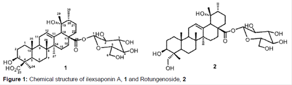
The compounds were identified as saponins (), by NMR and MS analysis. These compounds have already been reported [26-28].
Compound 1: 1H NMR (300 MHz, pyridine-d5): δ 1.09 (3H, d, J = 6.1 Hz), 1.27 (3H, s, CH3), 1.29 (3H, s, CH3), 1.43 (3H, s. CH3), 1.73 (3H, s, CH3), 1.77 (3H, s, CH3), 2.97 (lH, s, H-18), 3.30 (lH, dd, J = 4, 11 Hz, H-3), 5.21 (lH, br s, 19-OH), 5.61 (lH, br t, H-12), 6.29 (lH, d, J = 6.8 Hz anomeric H). 13C NMR (75 MHz, pyridine-d5): δ 38.4 (C1), 28.5 (C2), 77.2 (C3), 48.6 (C4), 56.4 (C5), 23.7 (C6), 32.4 (C7), 40.1 (C8), 46.8 (C9), 37.1 (C10), 24.0 (C11), 128.9 (C12), 138.2 (C13), 42.0 (C14), 28.3 (C15), 26.1 (C16), 47.2 (C17), 53.7 (C18), 72.6 (C19), 41.6 (C20), 26.2 (C21), 37.0 (C22), 180.1 (C23), 23.7 (C24), 11.6 (C25), 18.4 (C26), 25.5 (C27), 177.7 (C28), 26.5 (C29), 15.8 (C30), 94.7 (G-1), 74.8 (G-2), 77.6 (G-3), 70.1 (G-4), 78.0 (G-5), 61.6 (G-6). [M-H-] m/z 663.4648.
Compound 2: 1H NMR (300 MHz, pyridine-d5): δ 1.07 (3H, d, J = 6.1 Hz), 1.08 (3H, s, CH3), 1.09 (3H, s, CH3), 1.27 (3H, s. CH3), 1.41 (3H, s, CH3), 1.69 (3H, s, CH3), 2.98 (lH, s, H-18), 3.76 (2H, s, H23), 4.30 (lH, dd, J = 5.5, 11 Hz, H-3), 5.61 (lH, sapparent, H-12), 6.36 (lH, d, J = 7.5 Hz anomeric H). 13C NMR (75 MHz, pyridine-d5): δ 38.2 (C1), 26.0 (C2), 73.1 (C3), 41.9 (C4), 48.2 (C5), 17.9 (C6), 32.3 (C7), 39.8 (C8), 47.9 (C9), 36.5 (C10), 23.3 (C11), 128.1 (C12), 138.2 (C13), 41.5 (C14), 28.3 (C15), 25.1 (C16), 48.1 (C17), 53.6 (C18), 72.4 (C19), 41.3 (C20), 25.7 (C21), 36.9 (C22), 66.3 (C23), 11.3 (C24), 16.3 (C25), 15.2 (C26), 23.4 (C27), 177.2 (C28), 25.8 (C29), 14.9 (C30), 94.3 (G-1), 72.2 (G-2), 77.1 (G-3), 69.8 (G-4), 77.2 (G-5), 61.1 (G-6). [M+Na+] m/z 673.3937.
Antileishmanial and cytotoxic activities
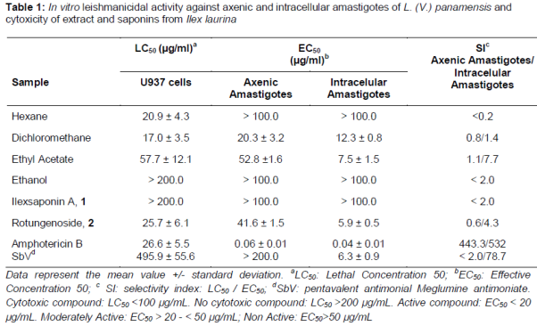
The leishmanicidal activity and cytotoxicity of extracts and saponins as well as meglumine antimoniate and amphotericin B, were evaluated following the method previously reported in the literature [21-25]. The leishmanicidal activity and cytotoxicity expressed as EC50 and LC50 values of extracts and saponins respectively, are shown in .
Based on the results shown in , the cytotoxicity of extracts varied among the solvent used. Thus, ethanolic extract was not cytotoxic for mammals U937 cells, exhibiting LC50 > 200 µg/mL. The remaining extracts using ethyl acetate, hexane and dichloromethane were cytotoxic with LC50 < 100 µg/mL. Amphotericin B was cytotoxic (LC50 = 26.6 ± 5.5 µg/mL) and meglumine antimoniate was not cytoxic (LC50 = 495.9 ± 55.6 µg/mL).
Leishmanicidal activity was observed for ethyl acetate and dichloromethane extracts against intracellular amastigotes with EC50 values < 20 µg/mL and moderately active against axenic amastigotes with values of EC50 > 20 µg/mL. The hexane (although cytotoxic for U937 cells) and ethanolic extracts were not active against intracellular or axenic amastigotes of L.(V.) panamensis with EC50 values > 100 µg/mL. Amphotericin B was highly active against both intracellular and axenic amastigotes of L. (V.) panamensis while meglumine antimoniate was also active against the intracellular form of the parasite.
Discussion
The activity observed in axenic amastigotes and intracellular amastigotes of ethyl acetate extract (EC50 < 60 μg/mL), suggests that this extract could be considered as promising in the search of new compounds against leishmaniasis. Based on this result, we performed a fractionation of the ethyl acetate extract and separated a major secondary metabolites which were identified as ilex saponin A, 1 and rotungenoside, 2 () by NMR and MS analysis [26-28].
Saponin 2 showed similar leishmanicidal activity and cytotoxicity to meglumine antimoniate and amphotericin B, respectively, drugs currently used for the treatment of leishmaniasis. In addition, the compound exhibited a selectivity index ≥1 (SI = 4.3) leading to greater activity against the parasite than the toxicity against the host cell. Some studies have shown that these compounds are active against various types of leishmaniasis [29-32]. Saponin 1 was not active or cytotoxic against L. (V) panamensis or U937 cells, respectively (). A possible explanation is the higher polarity due to carboxylic group, which could prevent the passage of the compound through the cell membrane.
The dichloromethane extract was the one that presented the best activity against axenic amastigotes of L. (V.) panamensis. Unfortunately, the cytotoxicity shown by this extract makes it a non-promising candidate in the search for new leishmanicidal compounds due to the risk of toxicity and no selectivity thereof. However, additional studies on cytoxicity using other cell lines are needed in order discriminate whether the cytoxicity showed by this extract is against tumoral or non-tumoral cells. The hexane extract showed activity against human cells but no activity against the Leishmania parasite and ethanolic extract was not potentially cytotoxic for the human U-937 cells but also was not active against L. (V.) panamensis; therefore, these extracts are not considered in the search for new compounds against cutaneous leishmaniasis.
Conclusion
Based on both leishmanicidal and cytotoxic activities, only ethyl acetate extract has potential to provide lead compounds for the development of new drugs to treat leishmaniasis. Two compounds have been isolated and identified in this study, both of which are saponins. Saponin 2 is a potential candidate for antileishmanial drug development based on its activity against Leishmania parasite. Although its cytotoxicity against U-937 cells is similar to amphotericin B, additional studies on cytotoxicity using other cell types are needed in order to determine whether the toxicity shown by this compound is against tumor or non-tumor cells. In addition, further studies are required to optimize the structure of the promising molecule and to validate if the in vitro activity against L. (V.) panamensis demonstrated here would also be observed in vivo.
Declarations
Acknowledgement
References
Archives


News Updates